


Philips
Philips Affiniti 70
Philips Affiniti 70
Couldn't load pickup availability


Description:
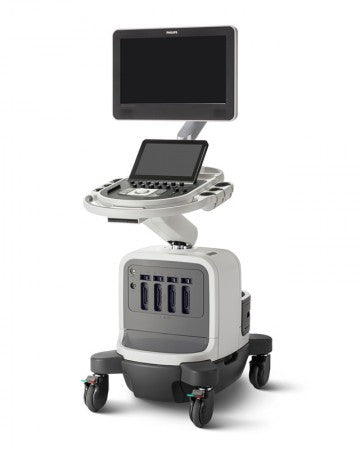
The Philips Affiniti 70, is a high-end shared service ultrasound machine that was developed in 2014 from the premium Epiq 7. The Affiniti 70 looks nearly identical to the Epiq 7 and 5 and offers many of the same features like single crystal PureWave transducers. The Affiniti 70 also has many of the same technologies as the Epiq series, including the amazing QLAB onboard quantification set and ShareWave elastography. The Philips Affiniti 70 is unique in the high-end price category in offering ShearWave. As a shared service ultrasound machine the Affiniti 70 excels at cardiology as well as womens health, vascular and radiology applications. Philips is most well known for its cardiac imaging superiority over other brands, but the Philips Affiniti 70 is also amazingly feature rich with excellent image quality at its price level. Doctors that like the Affiniti 70, but dont need the PureWave transducers or ShearWave Elastography should consider the less expensive Affiniti 50 instead.
Specifications
Transducers
Endocavitary C9-4v [ 4 – 9 MHz ] 128 elements, 10mmR, 181° field of view
Endocavitary C10-4ec [ 4 – 10 MHz ] 128 elements, 8mmR, 147° field of view
Bi-plane Endocavitary BP10-5ec [ 5 – 10 MHz ] 96 elements, 8.8mmR, 150° field of view
PureWave Endocavitary C10-3v [ 3 – 10 MHz ] 128 elements, 11.5mmR, 163° field of view
Convex C6-2 [ 2 – 6 MHz ] 128 elements, 10mmR, 63.7° field of view
Convex C8-5 [ 5 – 8 MHz ] 128 elements, 14mmR, 122° field of view
PureWave Microconvex C9-2 [ 2 – 9 MHz ] 192 elements, 45mmR, 102° field of view
PureWave Convex C5-1 [ 1 – 5 MHz ] 160 elements, 45mmR, 111° field of view
4D Convex V6-2 [ 2 – 6 MHz ] 192 elements, 55mmR, 100° x 85° volume field of view
4D Endocavitary 3D9-3v [ 3 – 9 MHz ] 128 elements, 26.1mm, 156° x 85° volume field of view
4D Linear VL13-5 [ 5 – 13 MHz ] 192 elements, 38.4mm, 38 mm x 30° volume field of view
Linear L18-5 [ 5 – 18 MHz ] 288 elements, 38.9mm, ultra-fine pitch
Intraoperative Linear L15-7io [ 7 – 15 MHz ] 128 elements, 23mm,
Linear L12-5 50 [ 5 – 12 MHz ] 256 elements, 50mm, fine pitch
Linear L12-3 [ 3 – 12 MHz ] 160 elements, 38mm, fine angle steering
Linear L12-4 [ 4 – 12 MHz ] 128 elements, 34mm, fine angle steering
Cardiac Sector S4-2 [ 2 – 4 MHz ] 80 elements, 5mm
PureWave Cardiac Sector S5-1 [ 1 – 5 MHz ] 80 elements, 20.3mm
Pediatric Cardiac Sector S8-3 [ 3 – 8 MHz ] 96 elements, 15.4mm
Neonatal Cardiac Sector S12-4 [ 4 – 12 MHz ] 96 elements, 9.78mm
Pediatric TEE transesophegeal S7-3t [ 3 – 7 MHz ] 48 elements, 5mm
xMATRIX TEE transesophegeal X7-2t [ 2 – 7 MHz ] 2,500 elements
Pedoff (CW Transducer) D5cwc [ 5 MHz ] Deep venous and arterial applications, non-imaging
Pedoff (CW Transducer) D2cwc [ 2 MHz ] Adult cardiology applications, non-imaging
Pedoff (PW Transducer) D2tcd [ 2 MHz ] Transcranial Doppler applications, non-imaging
Features
21.5” LCD display with four-way articulation
12” widescreen tablet-like touchscreen
4 transducer ports and one-handed transducer access
Intelligent software architecture
Combined 512 GB storage capacity
DVD+RW drive
Wireless networking
State-of-the-art ergonomic designed system cart for comfort and convenience
Easy-to-learn graphical user interface with reduced number of hard controls
Advanced control panel design with fewer, clustered controls
2D mode
Tissue Harmonic Imaging (THI)
M mode
Color Doppler
Color Power Angio Doppler
Pulse wave Doppler
Auto color and auto Doppler
Transport mode : The system lasts 45 minutes before recharge
PureWave crystal technology
Auto Doppler for vascular imaging
SonoCT real-time compound imaging
XRES adaptive image processing
iSCAN intelligent optimization
AutoSCAN intelligent optimization
SmartExam protocols
iOPTIMIZE intelligent optimization
Tissue aberration correction (TAC)
Coded beamforming
QuickSAVE feature
Cineloop review: up to 2,200 frames of 2D and color images
High Q automatic Doppler analysis
Imaging Nodes
B-mode
M-mode
(PW) Spectral Doppler
Steerable continuous wave (CW) Doppler
Tissue Doppler Imaging (TDI/TDI PW)
iRotate echo (X5-1 and X7-2t)
Live xPlane imaging
4D
Freehand 3D
Live 3D echo
STIC imaging
Panoramic imaging
Contrast imaging – cardiovascular, general imaging
Tissue Harmonic Imaging (THI)
Color Doppler
Color Power Angio imaging (CPA)
Strain-based elastography
Shear wave elastography
3D Panoramic imaging
Duplex mode
Triplex mode
Applications
Abdominal
Obstetrical
Fetal echo
Cerebrovascular
Vascular (peripheral, cerebrovascular, temporal TCD, and abdominal)
Abdominal vascular
Gynecological and fertility
Small parts and superficial
Musculoskeletal
Pediatric general imaging
Prostate
Echocardiography (adult, pediatric, fetal)
Stress echocardiography
Transesophageal echocardiography (adult and pediatric)
Surgical imaging
Interventional imaging
Contrast imaging
Bowel imaging
Strain elastography
Shear wave elastography (ElastPQ)
Perioperative
Epicardial echocardiography