


GE HealthCare
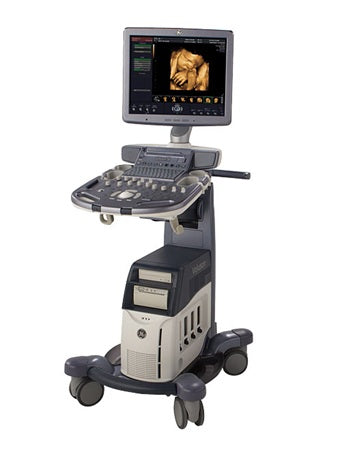
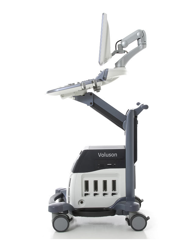
GE Voluson S8
GE Voluson S8
Couldn't load pickup availability

Description:
The GE ultrasound Voluson S8, is a high-end women’s health 4D ultrasound machine that has a price and features in-between the Voluson E8 and the Voluson S6. The Voluson line from GE is known worldwide for providing the best 4D imaging currently available though Samsung is catching up. The Voluson S8 and S6 look identical and even use most of the same transducers. The main difference is that the S6 uses half the channels of the Voluson S8. This produces a significant difference in image quality between the Voluson S8 and S6. The GE Voluson S8 uses the same cost effective RS transducers as the Voluson-i portable. The Voluson S8 offers near E8 level image quality at a lower price and much smaller, lighter form factor.
All Voluson ultrasound machines are focused on women’s health, especially obstetrics, gynecology, and fertility. Secondary applications include general imaging, adult & pediatric cardiology and neonatal cardiology. Doctors looking for shared service capabilities from a GE ultrasound machine should consider the GE Logiq S8 instead.
Specifications
Transducers
4D Convex RAB2-5-RS [ 1 – 5 MHz ] 192 elements, 46mm, max scanning depth 30cm
4D Convex RAB2-6-RS [ 2 – 6 MHz ] 192 elements, 46mm, max depth 26cm
4D Convex RAB4-8-RS [ 2 – 8 MHz ] 192 elements, 46mm, max scanning depth 26cm
4D Endocavitary RIC5-9W-RS [ 4 – 9 MHz ] 192 elements, 11.6mm, max scanning depth 16cm
Endocavitary E8C-RS [ 4 – 10 MHz ] 128 elements, 10.7mm, max scanning depth 16cm
Convex C1-5-RS [ 2 – 5 MHz ] 192 elements, 56.1mm, max scanning depth 30cm
Convex 4C-RS [ 2 – 5 MHz ] 128 elements, 60.0mm, max scanning depth 30cm
Convex AB2-7-RS [ 2 – 8 MHz ] 192 elements, 40.0mm, max scanning depth 28cm
Microconvex 8C-RS [ 4 – 10 MHz ] 128 elements, 10.7mm, max scanning depth 16cm
Linear 12L-RS [ 4 – 12 MHz ] 192 elements, 37mm FOV, max scanning depth 8cm
Linear 9L-RS [ 3 – 8 MHz ] 192 elements, 37mm FOV, max scanning depth 14cm
Linear Matrix ML6-15-RS [ 4 – 13 MHz ] 336 elements, 50mm FOV, max scanning depth 12cm
Cardiac sector 3Sc-RS [ 1 – 4 MHz ] 64 elements, 50° FOV, max scanning depth 24cm
Pediatric cardiac sector 12S-RS [ 5 – 11 MHz ]18mm footprint, max scanning depth 12cm
Pencil Transducer P2D [ 2 MHz ] CW Split Crystal
Features
19” High-Resolution TFT LCD Monitor
Innovative user interface with onscreen menus
3 Active Probe Port
3D/4D Mode
4D Biopsy
HD-Flow
Automatic Tissue Optimization
Coded Excitation (CE)
Coded Harmonic Imaging with Pulse Inversion Technology
Tissue Doppler
Advanced SRI
CrossXBeam CRI
SonoNT
B Mode only
B + Power Doppler Mode
B + CFM Doppler Mode
B + HD-Flow Mode
B + CRI
B + CRI + CFM
B + CRI + PD
B + CRI + HD-Flow
B + B-Flow
Focus & Frequency Composite (FFC)
Inversion mode
High Resolution Zoom
Pan Zoom
SonoRenderStart
Steering
Tomographic Ultrasound Imaging (TUI)
Virtual Convex
VOCAL
Wide Angle on endovaginal probes
Beta-View
Patient information database
Image Archive on hard drive
3D/4D data compression (lossy/lossless)
Real-time automatic Doppler calculations
OB Measurement, Calculations & Reports
GYN Measurement, Calculations & Reports
Vascular Measurement, Calculations & Reports
Cardio Measurement, Calculations & Reports
Abdominal Measurement, Calculations & Reports
Small-Parts Measurement, Calculations & Reports
Urology Measurement, Calculations & Reports
Pediatrics Measurement, Calculations & Reports
Musculoskeletal Measurement, Calculations & Reports
Neurology Measurement, Calculations & Reports
Multigestational Calculations and Fetal Trending
CINE Memory: 140MB; up to 7,000 frames, Dual/Quad Display, Review Loop and speed
Integrated HD (500 GB)
Imaging Nodes
2D-Mode
M-Mode
PW Doppler
High PRF Doppler Mode
Color Flow Doppler Mode (CFM)
Power Doppler Mode (PD)
Elastography mode
XTD-Mode
Contrast Agent Mode (Contrast)
M-Color Flow Modes (M/CF, M/HD-Flow, M/TD)
Volume Modes (3D/4D):
4D Real Time
4D Biopsy
Real Time Triplex
CW Doppler Mode (CW)
Anatomical M-Mode
B-Flow Mode (BF)
VCI-A (Advanced Volume Contrast Imaging)
VCI OmniView
STIC
3D Static
Applications
Abdominal
Small Parts
Obstetrics
Gynecology
Cardiology
Urology
Vascular
Peripheral Vascular
Pediatrics
Neurology
Musculoskeletal
Breast